Monitoring the response of bone metastases to treatment using MRI and PET
10 Sep 2014
A review and position statement of the EORTC Imaging Group
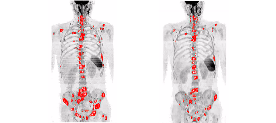
Imaging technologies are very useful in evaluating a patient’s response to cancer treatment, and this can be done quite effectively for most tumors using RECIST, Response Evaluation Criteria in Solid Tumors. However, RECIST works well for tumors located in soft tissue, but not so well for cancers that spread to the bone, such as is the case for prostate and breast cancers. More effort, therefore, is needed to improve our understanding of how to monitor the response of bone metastases to treatment using magnetic resonance imaging (MRI) and positron emission tomography (PET), and a recent EORTC Imaging Group review and position statement published in the European Journal of Cancer is a decidedly welcome contribution.
The paper published by these EORTC Imaging Group investigators highlights the most recent developments in MRI and PET and elucidates how these techniques can be used to detect bone metastases at an early stage as well as monitor the response of these to treatment. They describe the current state of the art in PET and MRI, the strengths, weaknesses and complementarity of various imaging techniques with respect to specific indications, and recommendations for choosing the most appropriate imaging technique.
Prof. Frederic Lecouvet of the Cliniques Universitaires Saint Luc in Brussels and lead author of this review and position statement says, “Assessing the response of metastases to treatment is something we do on a day to day basis, both in our oncology practice as well as for clinical trials. If the metastases occur in soft tissues, then we already have validated criteria, such as RECIST, that enable us to evaluate the response. A host of problems, however, limits our ability to measure the response to treatment of metastases found in bone. These range from the characteristics of bone metastatic disease, the structure of bone itself, to the sensitivity, specificity, and resolution of imaging methods available until now. We hope to resolve these issues, and our review is a step in the right direction.”
Techniques such as MRI and PET have the potential to detect the spreading of a cancer into the bone marrow at an earlier stage and also determine the extent of this spread, and the efforts of the EORTC researchers will go a long way towards making these techniques a routine part of oncology practice.
John Bean, PhD
EORTC, Medical Science Writer
Related News
EORTC: Advancing research and treatment for rare cancers
29 Feb 2024
EORTC Fellowship Programme: celebrating more than 20 years of impactful collaboration
22 Feb 2024
Appointment of Malte Peters as EORTC Strategic Alliance Officer
9 Feb 2024
Unique series of workshops in partnership with the European Medicines Agency (EMA)
7 Feb 2024
EORTC launches a prominent clinical trial in older patients with locally advanced (LA) HNSCC (Head and Neck Squamous Cell Carcinoma)
14 Dec 2023
Seven IMMUcan abstracts selected for ESMO Immuno-Oncology Congress 2023
6 Dec 2023
EORTC Quality of Life measures integrated in CDISC
20 Nov 2023
EORTC and Immunocore are collaborating to launch the ATOM clinical trial of tebentafusp in Adjuvant Uveal Melanoma
7 Nov 2023
Treatment with decitabine resulted in a similar survival and fewer adverse events compared with conventional chemotherapy in older fit patients with acute myeloid leukaemia
31 Oct 2023
New results and forthcoming EORTC trials in rare cancers, lung, head and neck, and breast carcinomas presented at ESMO 2023
20 Oct 2023